Cover Images
Why submit a cover image? Authors of accepted articles are strongly encouraged to submit an image to be considered for the cover of Diabetologia. This is a great opportunity to increase the visibility of your article as the cover image is publicised on our social media platforms and in table of contents alerts.
What are we looking for? We are open to your suggestions! Cover images can range from photographs to cartoons, single-cell micrographs to whole-tissue images. You may submit more than one image for consideration. See below for examples of previous cover images.
Requirements
- The suggested cover image must be linked to your accepted article — please provide a brief legend, indicating how the image is linked to your article, when submitting potential cover images.
- Authors must either hold the rights to the suggested cover image, or the copyright license must permit free reuse. Alternatively, if authors wish to solicit their own cover image or use a stock image, they are responsible for any fees associated with this.
- Images must be high resolution (≥300 dpi) and at least A4 size (210 × 297 mm) in portrait.
How to submit Please send potential cover images to the editorial office (diabetologia-j@bristol.ac.uk) as soon as possible after your manuscript has been accepted, quoting your manuscript number and title.
By submitting an image, authors agree to its use on social media platforms and in publicity material if the image is selected.

An immunofluorescence photograph of sensory neuron bodies within a dorsal root ganglion of a mouse model of type 1 diabetes (red, substance P; green, phospho-ribosomal protein S6 [P-rpS6])
Dang et al, https://doi.org/10.1007/s00125-019-4860-y
Cover credit: Elisa Avolio, Bristol Medical School, University of Bristol, Bristol, UK

The wide distribution of artificial lighting across China and the variety of sources of light exposure at night in modern society.
Zheng et al, https://doi.org/10.1007/s00125-022-05819-x
Cover credit: Shanghai Institute of Endocrine and Metabolic Diseases

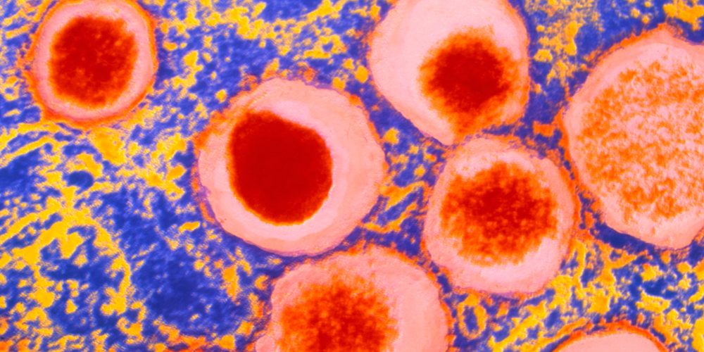
A scanning electron micrograph of extracellular vesicles appearing on the extracellular plasma membrane surface of a human islet cell.
Sims et al, http://dx.doi.org/10.1007/s00125-018-4712-1
Cover credit: Carmella Evans-Molina (Indiana University School of Medicine, Indianapolis, IN, USA)

The illustration, entitled ‘I want to create your pancreas’, is inspired by the popular Japanese anime movie ‘I want to eat your pancreas’. It depicts a researcher who vows to create a functional islet of Langerhans to save the life of a girl with severe type 1 diabetes.
Fukaishi et al, https://doi.org/10.1007/s00125-021-05560-x
Cover credit: Designed by Yoshio Fujitani, Gunma University, Gunma, Japan; illustrated by Hikoboshi (https://hikoboshi.wixsite.com/my-site); based on the artwork for ‘I want to eat your pancreas’, with permission from the publisher, Futabasha, Tokyo.

Illustration of a non-existent and improbable tool for clinical assessment.
Leslie et al, https://doi.org/10.1007/s00125-021-05471-x
Cover credit:
Concept: Michael E. J. Lean (University of Glasgow, Glasgow, UK).
Design: Lisa Juquet (https://lisajuquet.co/)

Drawing of a very small RNA molecule inside a crystal ball.
Garavelli et al, https://doi.org/10.1007/s00125-020-05237-x
Cover credit: Pencil/ink/digital drawing by Antonella Ficarra (www.containerstudio.it; www.behance.net/antofic)

An original charcoal, coal and wax drawing depicting a growing fetus in utero, with lipoproteins of differing sizes superimposed electronically on surrounding maternal tissue.
White et al, http://dx.doi.org/10.1007/s00125-017-4380-6
Cover credit: Original drawing and overall image design by Sara Spizzichino (www.saraspizzichino.com), incorporating lipoproteins designed by Antti Kangas of Nightingale Health Ltd (www.nightingalehealth.com)

An electron micrograph of a muscle biopsy obtained from a young and physically active individual with type 1 diabetes. The structural properties of mitochondria (red) and quantity of autophagy-associated structures in the subsarcolemmal (purple highlighting) and intermyofibrillar (highlighted in cyan) regions are visible.
Monaco et al, https://doi.org/10.1007/s00125-018-4602-6
Cover credit: Christian Lamberz (German Center for Neurodegenerative Diseases [DZNE], Bonn, Germany), Thomas Hawke (McMaster University, Hamilton, ON, Canada) and Christopher Perry (York University, Toronto, ON, Canada).

